
PET-CT vs. MRI for Cancer Screening
PET/CT and MRI don’t answer the same question, and for someone who feels fine, neither takes the place of proven organ-specific screening. When you do have symptoms, an abnormal result, or a cancer diagnosis, the next scan should be picked around that specific clinical question — not the other way around.
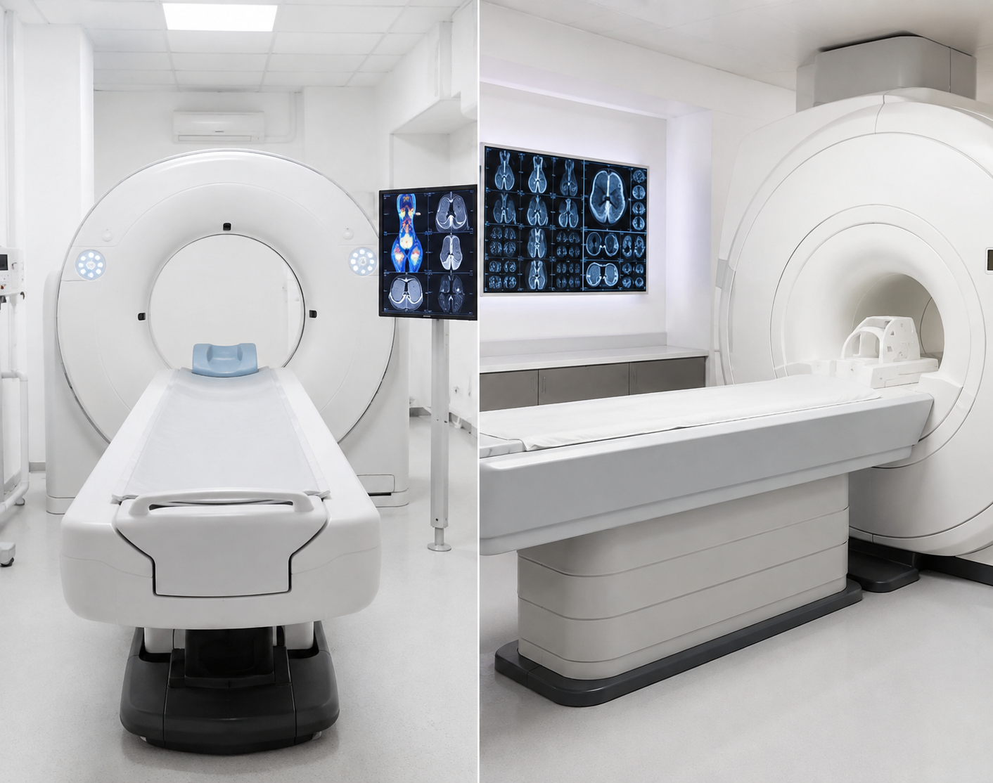
For average-risk adults with no symptoms, PET/CT and whole-body MRI are not substitutes for evidence-based cancer screening. PET/CT is mainly an oncology tool for selected cancers; MRI is strongest when detailed soft-tissue imaging is needed. A normal result from either test does not rule out every cancer.
Use the scan to answer a defined question, not as a universal cancer check.
The same scan means different things in different situations
Screening and diagnosis aren’t the same thing — and they shouldn’t be handled that way.
Begin with proven age- and risk-based screening. Japan’s National Cancer Center does not recommend PET as a replacement for the five population screening programs with established evidence of reducing cancer deaths.
This is a diagnostic problem, not routine screening. The useful next test depends on the organ, symptom and previous finding. Targeted CT, MRI, ultrasound, endoscopy, laboratory testing or biopsy may be more appropriate than a whole-body scan.
PET/CT or MRI may be used for staging, treatment planning, response assessment or suspected recurrence depending on the cancer type. At this point, imaging should follow an oncology plan rather than a general checkup package.
The original cancer type, pathology, stage, treatment dates and earlier imaging usually matter more than choosing a scan by name.
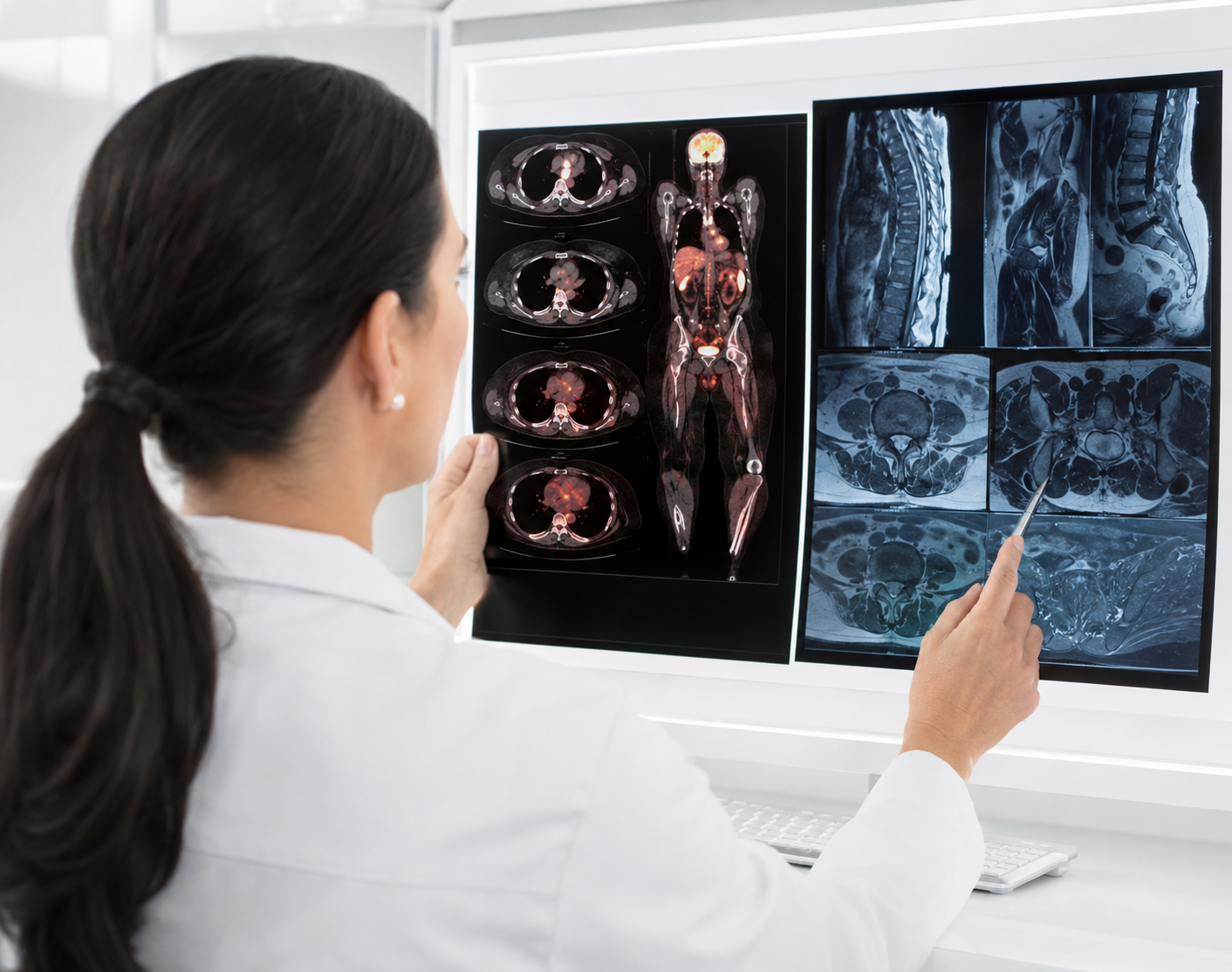
What each test can and cannot answer
FDG uptake plus CT anatomy.
High-detail tissue anatomy; some protocols add diffusion or contrast.
Not an evidence-based replacement for established population screening.
ACR says evidence is insufficient for total-body MRI in asymptomatic people without relevant risk factors.
Yes. Radiation comes from both the radioactive tracer and CT.
No ionizing radiation.
Often used for staging, response or recurrence when the cancer and tracer are suitable.
Often used for local detail in the brain, liver, pelvis, prostate and other soft tissues.
Inflammation and other benign processes can take up FDG; small or low-uptake tumors may be missed.
Non-specific incidental findings can lead to further imaging or procedures.
No. Suspicious uptake may still need tissue sampling or another targeted test.
No. Suspicious imaging findings may still need biopsy or other confirmation.
Numbers that change how the result should be interpreted
PET/CT dose and preparation change from one protocol to the next. These numbers come from published references — they’re not a single fixed dose or a promise that every facility works the same way.
Radiation dose estimates exposure — it doesn’t directly predict your personal cancer risk. Protocol, body size and CT settings all play a role.
Already have a package quote but you’re not sure what it covers?
Send the included tests and scan protocol — we can help you compare the scope before you book.
Start with the test proven for that organ
Japan’s National Cancer Center points to five population screening programs backed by established evidence. PET, CT and tumor markers may matter after a finding or diagnosis — but they’re not meant to take the place of these programs.
Source: National Cancer Center Japan cancer screening guidance.
Screening tests can have false-negative results, so stick with the age- and risk-based schedule that applies to you unless a qualified clinician tells you otherwise.

Details that can change the scan or the preparation
Send these details early — they can shift whether PET/CT, MRI, another test or a different prep plan makes more sense.
Not sure which records to pull together before asking for a quote?
Start with your age, main concern, previous test results and preferred dates in Japan.
Compare the scope before comparing the price
A PET/CT scan isn’t the same product as a head MRI, and neither is the same as a whole-body MRI protocol or a full health-check package. A low headline price may leave out specialist review, contrast, extra imaging or international support — so compare the scope before you compare the number.

Public price examples checked 11 August 2026. Sources: Fujimoto General Hospital and Nihonbashi Muromachi Mitsui Tower Midtown Clinic. Prices and visitor fees may change.

A scan finding is not the same as a cancer diagnosis
Continue the screening schedule that matches your age and risk. One negative whole-body scan cannot guarantee that every cancer is absent.
Ask which organ is involved, how concerning the finding is and which targeted follow-up test is recommended.
Ask what needs confirmation. Depending on the organ, the next step may be targeted CT or MRI, ultrasound, endoscopy or biopsy. A Japanese PET/CT patient guide notes biopsy is often required for a definitive cancer diagnosis.
Staging and treatment planning become cancer-specific. PET/CT or MRI may then be selected according to tumor type, stage and the question that needs to be answered.
A PET/CT or MRI result alone cannot give a reliable survival estimate. Prognosis depends on the confirmed cancer type, stage, tumor biology, treatment options and overall health.
Japan health-check planning and coordination
We help international visitors pull together their medical information, understand what a proposed check actually includes, and coordinate the practical steps in Japan. We aren’t a hospital or clinic.
Medical suitability, diagnosis and treatment decisions remain with qualified medical professionals.

Age, main concern, symptoms, family history, previous reports, cancer history if relevant and preferred dates.
We separate routine screening from diagnostic follow-up and identify what needs clarification.
We check the included tests, preparation, exclusions and possible extra fees.
We coordinate appointment communication, documents, timing and language support.
You know what is booked, what to bring, how to prepare and what needs follow-up.
Use the guide that matches your next decision
Questions patients usually need answered first
The right answer changes with symptoms, age, smoking history, family history, previous cancer and earlier test results.

Which is better for cancer screening, PET-CT or MRI?
Neither is universally better. For average-risk adults without symptoms, proven organ-specific screening should come first. PET/CT is mainly used for selected oncology questions; MRI is chosen when detailed soft-tissue imaging is needed.
Can PET-CT detect every cancer?
No. Small or low-FDG-uptake tumors can be difficult to detect, while inflammation can also look active. A Japanese hospital guide notes that lesions below 1 cm become harder to detect as size decreases.
Does a normal whole-body MRI rule out cancer?
No. ACR does not recommend total-body MRI as routine screening for asymptomatic people without relevant risk factors, and MRI does not replace established organ-specific tests.
How much radiation is in PET-CT?
There is no fixed dose. RadiologyInfo lists about 22.7 mSv for a typical adult whole-body protocol. IAEA gives about 8 mSv for the PET component, while CT dose changes with the CT protocol.
Should I do PET-CT and MRI together for maximum coverage?
Not automatically. More imaging can produce non-specific findings, extra follow-up and higher cost. The useful combination depends on the organ and clinical question.
What if I already have symptoms or an abnormal result?
That is a diagnostic question, not routine screening. The next test should target the symptom or abnormal organ rather than simply adding a whole-body scan.
What should I send before asking for a quote?
Send your age, main concern, symptoms, family history, previous reports, cancer history if relevant, diabetes status, implants or devices and preferred dates in Japan.
Evidence and price sources
Reviewed 11 August 2026. Dose, screening recommendations, facility protocols and prices should be rechecked before use.
Tell us what you want to check and what you already know
Send your age, main concern, previous results and preferred dates in Japan. We’ll help you organize the next questions and compare the practical options before you commit to a package.
Educational and coordination information only. We aren’t a hospital or clinic and don’t provide diagnosis or treatment. If you have persistent or concerning symptoms, seek diagnostic medical evaluation rather than waiting for routine screening.