
- You may not need a new MRI if your diagnosis is already clear and your current images still match your symptoms.
- You may need MRI if the doctor still needs to identify the source of the problem or decide whether treatment is suitable.
- You may need another type of scan if X-ray, CT, or ultrasound can answer the question better.
Before booking another scan, ask one practical question: “What MRI finding could change my diagnosis, treatment target, or eligibility?”
The information below mainly applies to regenerative and cell-based treatment for joints, the spine, chronic pain, and related conditions. Hematopoietic stem cell transplantation for leukemia and other blood diseases uses a different pre-treatment work-up.
Japan Does Not Require MRI for Every Patient
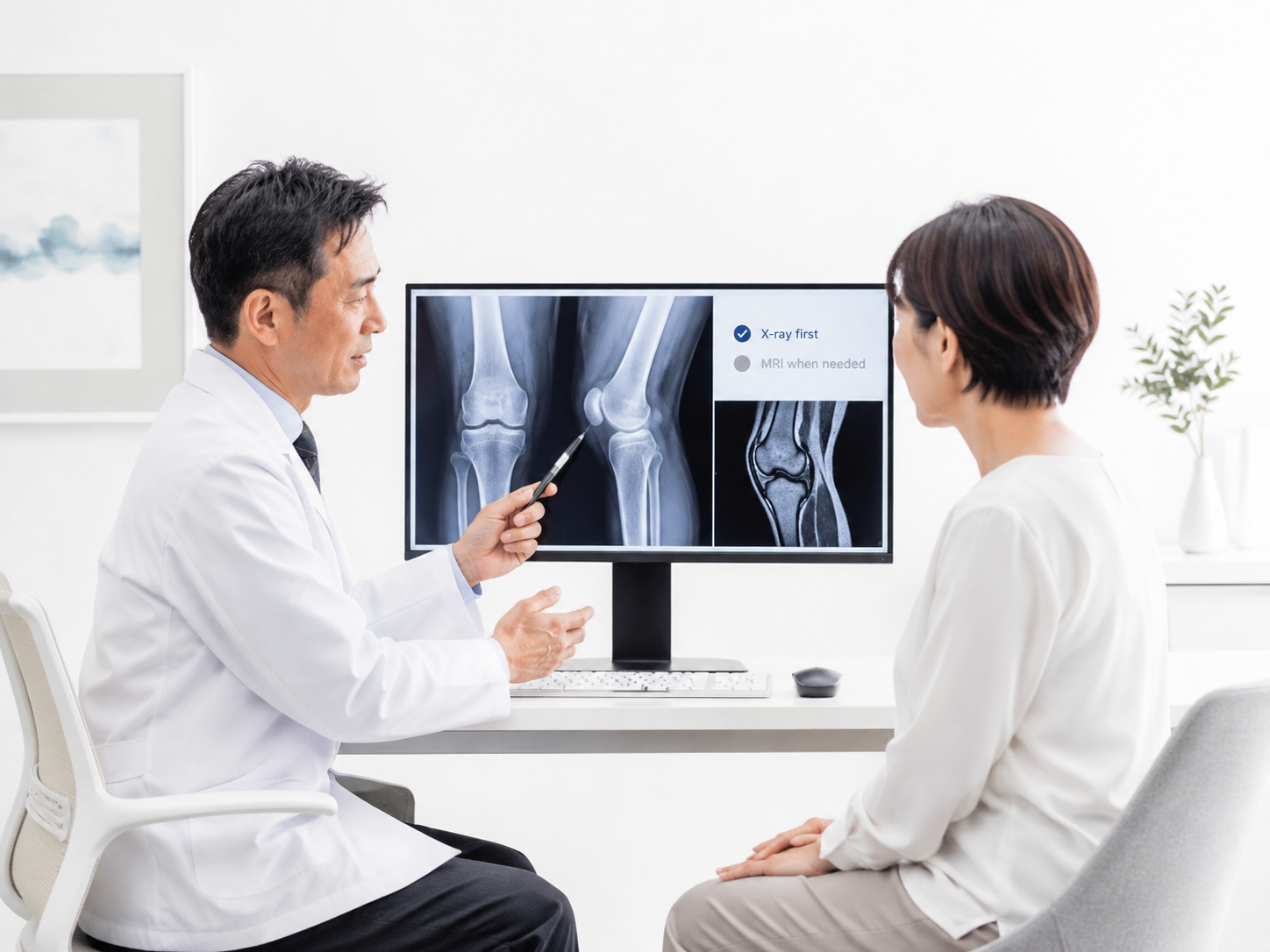
There is no Japanese rule that makes MRI compulsory for every regenerative medicine patient.
Under the Act on the Safety of Regenerative Medicine, covered regenerative medicine is divided into three risk classes. The review, submission, reporting, and record-keeping requirements differ by treatment.[1]
A clinic can still require MRI as part of its own medical protocol. That is a treatment-specific medical decision, not a nationwide rule.
For regenerative medicine in Japan, the imaging should match the condition being treated, the symptoms, the body area involved, the scans already available, and the procedure under consideration.
Use MRI Only When It Can Change the Decision
A useful MRI should give the doctor information that matters to the next step. In practice, that usually means one of four things:
- Confirm the diagnosis: work out whether the problem involves cartilage, a meniscus, tendon, ligament, disc, nerve, bone, or another structure.
- Find another problem: detect a fracture, osteonecrosis, infection, tumor, major tear, nerve compression, or another condition that may need different treatment.
- Choose the treatment target: show where the damage is and how much tissue is involved.
- Provide a baseline: give doctors a valid pre-treatment image when later imaging has a clear clinical or research purpose.
If the scan would not change the diagnosis, treatment target, or treatment plan, repeating it may not add much value.
Knee Pain Usually Starts With an X-Ray
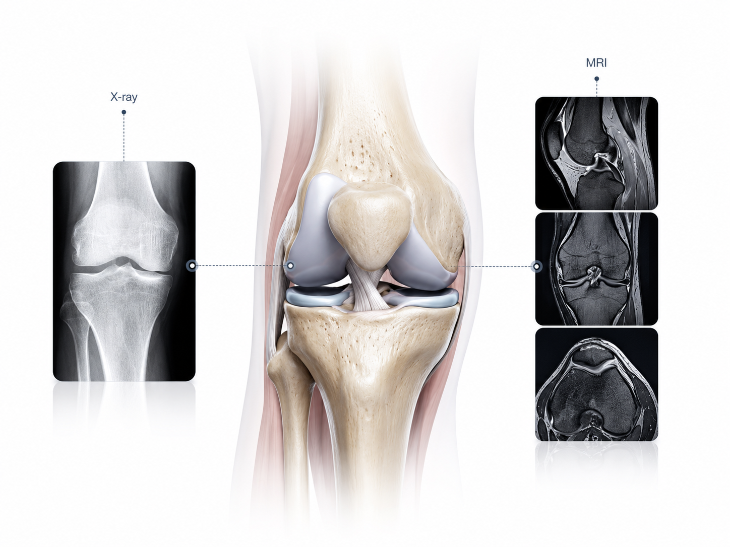
For chronic knee pain, the American College of Radiology (ACR) rates knee radiography as Usually Appropriate for initial imaging. MRI without contrast is rated Usually Not Appropriate as the routine first test for chronic knee pain.[2]
Weight-bearing knee X-rays can show:
- loss of joint space;
- bone spurs;
- bone shape;
- joint alignment;
- advanced osteoarthritis;
- some previous fractures and bone changes.
Knee MRI can show more detail in:
- cartilage;
- menisci;
- ligaments;
- tendons;
- muscles;
- bone marrow;
- joint fluid;
- other soft tissues.
MRI does not use ionizing radiation and can provide detailed images of both bone and soft tissue.[3]
For someone looking at regenerative treatment for knee joint disorders, MRI is not automatically the better test simply because it gives more detail. X-ray and MRI answer different questions.
Get a Knee MRI When the X-Ray Is Not Enough
Knee MRI is more useful when:
- the X-ray does not explain persistent symptoms;
- there is unexplained swelling or joint fluid;
- the knee repeatedly catches or locks;
- there has been a new injury;
- a meniscus or ligament injury is suspected;
- cartilage or the bone beneath it needs closer assessment;
- there has been previous cartilage or meniscus surgery;
- another condition besides ordinary osteoarthritis is suspected.
If chronic knee pain continues and the initial X-ray is negative or shows joint fluid, ACR rates MRI without IV contrast as Usually Appropriate for the next examination. If the X-ray already shows degenerative changes, MRI without contrast is rated May Be Appropriate rather than automatically necessary.[4]
Take a patient whose standing X-rays already show severe joint-space loss, bone spurs, deformity, and symptoms typical of advanced osteoarthritis. MRI may show the joint in greater detail, but it may not change the diagnosis.
The situation is different when the X-ray looks only mildly abnormal but the patient has major swelling, locking, or loss of function. In that case, MRI may uncover a separate problem that changes what the doctor recommends.
An Abnormal Knee MRI Does Not Always Explain the Pain
Many people have abnormal knee MRI findings even though their knees do not hurt.
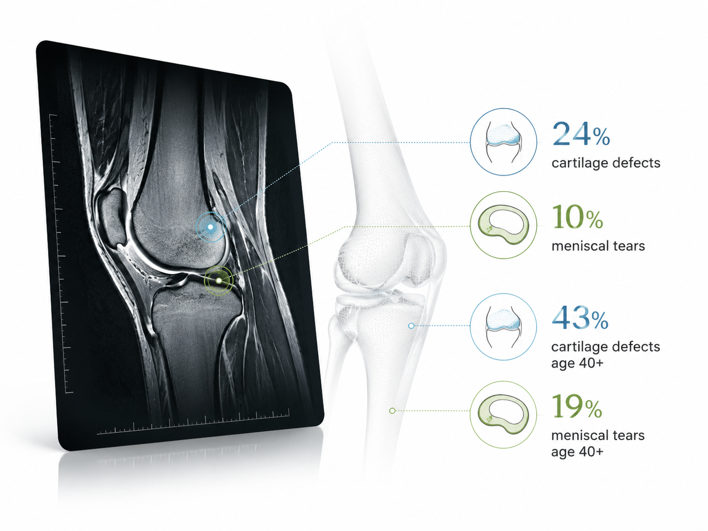
A systematic review of 63 studies examined 5,397 knees from 4,751 adults who had no knee symptoms and no previous knee injury. MRI still found:
- cartilage defects in about 24% of knees;
- meniscal tears in about 10%.
Among symptom-free adults aged 40 or older, cartilage defects were present in about 43% and meniscal tears in about 19%.[5]
So a meniscus tear or cartilage defect on MRI is not enough, by itself, to explain why a knee hurts. The doctor still needs to compare the scan with the patient’s symptoms, physical examination, movement problems, and day-to-day function.
Severe MRI Damage Does Not Predict a Better Stem Cell Result
More visible damage does not mean a patient is more likely to respond to cell-based treatment.
- Imaging severity: how damaged the joint looks.
- Symptom severity: how much pain, stiffness, or loss of function the patient has.
- Treatment response: how much the patient improves after a particular treatment.
These are three separate things. One patient may have severe cartilage loss but only moderate symptoms. Another may have substantial pain even though the MRI looks less dramatic.
For knee treatment, doctors may also need to consider body weight, leg alignment, joint stability, range of motion, previous surgery, other medical conditions, previous treatment, and the patient’s goals. MRI alone cannot decide stem cell treatment eligibility.
Shoulder and Hip MRI Should Affect the Plan
In the shoulder, MRI can help tell the difference between a smaller tendon injury and a full-thickness rotator cuff tear, tendon retraction, muscle loss, labral damage, or advanced joint disease. Those findings can lead to very different treatment choices.
Hip MRI can help identify cartilage damage, labral injury, tendon problems, bone-marrow abnormalities, osteonecrosis, and some fractures that are hard to see on an initial X-ray.
If the MRI result would not change the treatment decision, another scan may not be useful.
Simple Back Pain Does Not Automatically Need MRI
Routine lumbar MRI is not recommended for every patient with uncomplicated back pain.
ACR rates lumbar MRI without contrast as Usually Appropriate when symptoms remain persistent or progressive after about six weeks of appropriate treatment and the patient may be a candidate for surgery or another intervention.[6]
MRI may be needed sooner when the doctor is concerned about serious nerve compression, infection, cancer, fracture, or another urgent problem.
A Disc Bulge Does Not Automatically Cause Back Pain
Spine MRI often shows age-related changes in people who have no symptoms at all.
A systematic review of 33 studies involving 3,110 people without symptoms found:
- disc degeneration in about 37% of 20-year-olds and 96% of 80-year-olds;
- disc bulges in about 30% of 20-year-olds and 84% of 80-year-olds.
The authors concluded that many degenerative findings seen on spine imaging are part of normal aging and must be read together with the patient’s clinical condition.[7]
A report that says “disc bulge” therefore does not prove the disc is the source of pain.
The doctor should check whether the MRI matches:
- the location of pain;
- pain traveling into an arm or leg;
- numbness or weakness;
- the neurological examination;
- other possible causes.
This matters when assessing cell-based treatment for chronic pain, because a scan can support a diagnosis without proving that a particular treatment will help.
Neurological Conditions Need Their Own Imaging Plan
Spinal cord injury, stroke, multiple sclerosis, traumatic brain injury, and neurodegenerative diseases do not use one shared MRI protocol.
MRI may be used to locate an injury, identify compression or inflammation, rule out another diagnosis, or provide a baseline.
An abnormal brain or spinal cord MRI does not show that a cell treatment can restore lost neurological function. The evidence still has to match the specific disease, cell product, dose, treatment route, and patient group.
IV Cell Treatment May Need Other Tests Instead
When cells are given intravenously rather than injected into a specific joint or spinal structure, MRI may have little direct role in the procedure.
Depending on the disease and treatment, the work-up may instead include:
- complete blood count;
- kidney function;
- liver function;
- blood-clotting tests;
- infection screening;
- cardiovascular assessment;
- disease-specific tests.
The tests should fit the diagnosis and treatment risks. A patient does not need MRI simply because cells are part of the treatment.
Use an Old MRI When It Still Matches Your Condition
There is no universal rule that an MRI becomes unusable after three months, six months, or one year.
An existing MRI may still be adequate when:
- it covers the correct body part;
- the images are good quality;
- the required structures are visible;
- your symptoms have not changed significantly;
- you have not had a major new injury;
- you have not had surgery since the scan.
A 10-month-old MRI can still be useful for stable chronic knee arthritis. A two-month-old MRI may already be outdated after a new fall, new weakness, major swelling, or an operation.
If you already have scans, send them before paying for another examination. The stem cell treatment process in Japan may start with a review of existing imaging, diagnosis reports, treatment history, and other records before additional testing is requested.
Send the DICOM Images, Not Just the Report
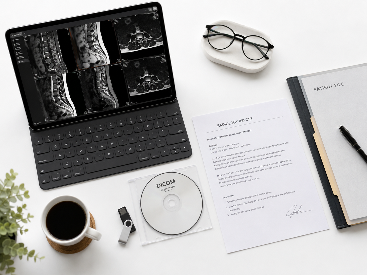
Whenever possible, send both the radiology report and the original MRI files.
MRI images are commonly stored as DICOM files. The report is a written summary; the DICOM files let another physician review the complete scan.
A report that says “moderate cartilage loss” may not tell the treating doctor:
- the exact location of damage;
- the size of the damaged area;
- whether the meniscus is involved;
- whether the bone under the cartilage is abnormal;
- whether other parts of the joint are also affected.
Before traveling, confirm that the physician making the treatment decision has reviewed the actual images, rather than only a PDF report received by administrative staff.
Most Routine Knee MRI Does Not Need Contrast
Most routine MRI examinations for knee osteoarthritis and common meniscal or ligament problems do not need IV contrast.
In the common chronic knee-pain situations where MRI is appropriate, ACR generally recommends MRI without IV contrast. MRI with and without contrast is rated Usually Not Appropriate in the routine scenarios covered by the guideline.[8]
Contrast may still be useful for selected problems such as suspected infection, tumor, certain inflammatory conditions, or some postoperative complications.
MRI contrast commonly contains a gadolinium-based agent. It is different from the iodine-based contrast widely used for CT scans.[9]
Contrast is not an automatic upgrade. It should be used when the doctor needs it to answer a specific question.
Metal and Implants Do Not Automatically Rule Out MRI
Tell the imaging center if you have a pacemaker, cochlear implant, nerve stimulator, medication pump, aneurysm clip, metal fragments, joint replacement, spinal hardware, or another implanted device.
Some devices are MR Conditional, which means MRI can be performed only under specified conditions. The imaging team may need the device name, manufacturer, model, and implant information before allowing the scan.[10]
Metal can also distort the image around an implant. Depending on what the doctor needs to see, a modified MRI technique, CT, X-ray, or another imaging method may be more useful.
If MRI Is Not Possible, Use Another Test
Patients who cannot complete a conventional MRI may still be assessed with:
- X-ray;
- CT;
- ultrasound;
- a different MRI setup;
- another disease-specific investigation.
The alternative should answer the same medical question the MRI was supposed to answer.
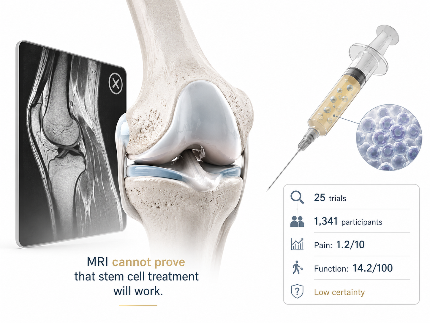
MRI Cannot Prove That Stem Cell Treatment Will Work
An MRI can show cartilage loss, tendon damage, disc degeneration, or other structural changes. It cannot prove that injected cells will:
- grow normal new cartilage;
- reverse osteoarthritis;
- repair a damaged disc;
- permanently remove pain;
- prevent future surgery;
- restore a joint to its previous condition.
Those questions have to be answered with clinical evidence from the specific treatment and condition.
Knee Stem Cell Evidence: 25 Trials and 1,341 Participants
A 2025 Cochrane review included 25 randomized trials and 1,341 participants with knee osteoarthritis. Eight trials with 459 participants compared stem cell injections with placebo injections.[11]
Compared with placebo, the review found:
- Pain: 7 studies with 445 participants showed an average improvement of 1.2 points on a 0-to-10 scale at up to six months.
- Function: 7 studies with 432 participants showed an average improvement of 14.2 points on a 0-to-100 scale at up to six months.
Cochrane rated the evidence for pain and function as low certainty. Trials used different cell sources, doses, and preparation methods. The review also found uncertainty about quality of life, overall treatment success, structural progression, and important safety outcomes.[12]
These results suggest that some patients may have modest symptom improvement. They do not prove that an arthritic knee has been structurally rebuilt.
Do Not Use Before-and-After MRI as Proof of Cartilage Regrowth
Two MRI screenshots are not enough to prove that normal cartilage has grown back.
If before-and-after MRI is being used to support a regeneration claim, check:
- whether the same MRI protocol was used;
- whether exactly the same area was compared;
- whether cartilage was measured rather than judged only by appearance;
- who interpreted the scans;
- whether the reader was independent of the treatment provider;
- whether pain and function also improved.
A patient can have less pain without cartilage regrowth. An MRI can also look different without producing a meaningful improvement in walking, function, or quality of life.
Japan Has Three Different Regulatory Situations
Patients should keep these three situations separate.
Regenerative medicine provided as treatment: Covered non-research treatment follows the applicable procedures under the Act on the Safety of Regenerative Medicine.
Regenerative medicine conducted as research: MHLW states that covered regenerative medicine research uses Japan’s jRCT system for submission and public registration.[13]
Approved regenerative medical product: This follows a separate product-regulation process. PMDA publishes review information for regenerative medical products that have received marketing approval in Japan.[14]
A submitted regenerative medicine provision plan does not mean the exact therapy has received marketing approval as a regenerative medical product.
The distinction is explained in more detail in Japan regenerative medicine registration and compliance.
If a provider uses the word “approved,” ask for:
- the exact treatment or product name;
- the disease or indication;
- the regulatory route;
- whether it is treatment, research, or an approved regenerative medical product.
Know the Exact Cell Treatment Before Planning Around MRI
“Stem cell therapy” is not a precise treatment name.
Before deciding whether the available imaging is enough, identify:
- the cell type;
- the source of the cells;
- whether the cells are your own or from a donor;
- whether they are cultured or otherwise processed;
- the route of administration;
- the planned number of treatments.
The differences between autologous and allogeneic stem cells matter because cell source, processing, immune considerations, and evidence are different.
When cells are cultured or processed, documentation from the cell processing center deals with manufacturing and quality-control questions that MRI cannot answer.
The International Society for Stem Cell Research advises patients considering unproven stem cell interventions to check the exact treatment, scientific evidence, expected results, risks, and oversight rather than relying on testimonials or broad claims.[15]
Get These Answers Before Flying to Japan
Before paying for non-refundable travel, send the records you already have:
- your diagnosis or specialist report;
- current medications;
- previous treatment history;
- operation reports;
- X-rays;
- MRI or CT reports;
- original DICOM images;
- recent laboratory results when relevant.
The medical travel guide for stem cell treatment in Japan covers the records and practical checks that may be needed before international travel.

Before paying for treatment or another MRI, get clear answers to these questions:
- What is my exact diagnosis?
- Why is a new MRI needed?
- Can my existing DICOM images be used?
- What MRI finding could change or cancel treatment?
- Has the treating physician reviewed my original images?
- What exact cell treatment is being considered?
- What human evidence supports it for my diagnosis?
- What is its regulatory status in Japan?
These MRI Practices Deserve More Questions
- Eligibility is confirmed before a physician reviews the diagnosis and imaging.
- No one can explain why MRI is needed.
- A sales coordinator interprets the MRI and tells the patient treatment is necessary.
- An abnormal scan is used as proof that stem cells will regenerate tissue.
- No possible MRI result would change or cancel the treatment.
- The provider says “government approved” but cannot identify what is actually approved or registered.
Seek Urgent Care for These Symptoms
Do not wait for treatment planning abroad if you develop:
- new loss of bladder control;
- new loss of bowel control;
- numbness around the groin or inner thighs;
- rapidly worsening arm or leg weakness;
- major new neurological problems;
- severe symptoms after significant trauma;
- symptoms suggesting a serious infection.
ACR rates lumbar MRI as Usually Appropriate when cauda equina syndrome is suspected and uses different imaging pathways when cancer, infection, or other serious disease is a concern.[16]
Finally
You do not need a new MRI simply because you are considering stem cell treatment in Japan. For chronic knee pain, X-ray is usually the first test, while MRI becomes more useful when the X-ray leaves an important question unanswered. MRI findings also need to be read carefully: a review of 5,397 symptom-free knees found cartilage defects in 24% and meniscal tears in 10%. Treatment results are a separate issue. A 2025 Cochrane review of 25 randomized trials and 1,341 knee-osteoarthritis patients found only low-certainty evidence of modest average improvements in pain and function. Before repeating MRI, ask what result could actually change your diagnosis, treatment target, or eligibility.